

Photography enthusiasts know that light is the true designer of the face. So much so that Hollywood stars have their own personal “illuminator”, which I impose on every director, to protect their beauty in front of the camera.
What is often not known is that in addition to the light that “illuminates”, there is also the light that signals what is not evident to the naked eye. For example, the famous X-rays, which reach the internal organs and describe their physiological confirmation or not, the classic radiographs. Even infrared light reveals what is invisible to the eye at night. You know the war movies?
In the cosmetic field, there is a particular type of light that reads the skin and reveals its superficial and/or deep marks, which demonstrates how much a cosmetic is capable of accomplishing in a given time, thanks to the measurement of the mark’s quality and quantity before and after treatment.
For MyCli, we use the VISIA® skin analysis system, an innovative and precise scanner for computerized face analysis that uses three different types of light: cross-polarized, UV, and Canfield’s RBX® technology. The patients put their face in front of the machine and in a few seconds a beam of light “reads” its surface.
Subsequently, a dedicated software analyzes the images, transforms them into numerical values, and allows objective monitoring of the cosmetic treatment’s progress over time.
To evaluate the benefits of a cosmetic protocol it is necessary to repeat the same analysis after a certain time interval. But what is the best control time? It depends on the tested product and what it contains. If the product promises immediate effect, then the images will be taken 30 minutes or two hours apart. In the case of products or protocols that require more time to act, we will proceed with the acquisition of images after 2 weeks or 28 days.
The parameters we measured for some MyCli protocols are:
- Spots and discolorations. Signs of evident discoloration on the surface of the skin, normally visible to the naked eye. In this case, we obtain not only the two images, before and after the treatment, but also the calculation of the improvement expressed as a percentage.
- Wrinkles. Differentiation of light wrinkles from deep ones, associated with a decrease in skin elasticity. Here the scanner reveals whether after the protocol the depth of the wrinkles has decreased and by how much.
- Skin texture. Analysis of the uniformity of the skin’s smoothness, highlighting “depressed” and “raised” areas that indicate variations on the skin surface. If after the treatment the uniformity increases, it will be a sign of much brighter, smoother, and more “perfect” skin.
- Pores. Analysis of the superficial circular openings of the sweat gland ducts. Very useful for verifying the effectiveness of exfoliants and protocols for oily or asphyxiated skin.
- Deep UV-caused spots. Analysis of the irregular color areas that are under the skin surface, the spots that are not seen but sooner or later will arrive. This measurement is essential in evaluating the effectiveness of stain protection procedures.
- Brown spots. Signs of pigmentation and discoloration above and below the skin surface, the spots already present, and their progressive depigmentation.
- Porphyrin deposits. Bacterial excretions that can deposit in the pores and demonstrate how an antibacterial product can fight phenomena such as acne or rosacea.
- Red areas. Detection of the presence of blood vessels under the skin surface, often associated with inflammation that as they decrease indicates to us how soothing procedures work.
Therefore, it is possible to accurately assess the state of any person’s skin. The data obtained will actually quantify the number of alterations present on the skin and will subsequently be compared with a database containing data relating to a population homogeneous for age, phototype and sex.
In the next points, there are other forms of effectiveness evaluations. See you soon!
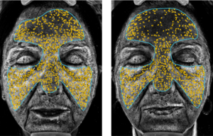
Here is an analysis carried out before (left photo) and after (right photo) using the MyCli procedure for spots and uneven complexion.

Dr. Alessandra Gava
Pharmaceutical Biotechnology Specialist